Robotic partial Nephrectomy
Education event Sponsored by: Intuitive Surgical
Conference Name: Live Workshop on Robotic partial Nephrectomy
Conducted by: Intuitive Surgical & Dr. Yajvender Rana
Surgeon(s)/Speaker(s): Dr. Yajvender Rana
Surgical Procedure: Kidney-Robotic Radical Nephrectomy
Location: BLK MAX Hospital New Delhi
Indexsteps
1. Placing French Foley catheter : A 16-French Foley catheter was placed under all aseptic precaution.
2. Donar details :
3. Anasthesia to be given to Donar and Recipient :
4. Role of robot in kidney transplant :
5. Port placement in Recipient :
6. Marking of ports and its placement :
7. Hand controls on the robot :
8. Foot controls on Robot :
9. Robotic buccal graft urethropathy :
10. Robotic buccal graft ureteroplasty :
11. position and port placement :
12. Urolift system :
13. Q and A :
14. Q and A :
15. Q and A :
16. Q and A :
17. Q and A :
18. Completion of Uretic reimplantation :
19. Q and A :
20. Genitalia and abdomen prepping and draping : The area of genitalia and abdomen were prepped and draped in a standard fashion.
21. Port insertion : Under direct vision, the 8 mm robotic trocars were placed, one in subcostal and one in the lower quadrant. Making an appropriate triangulation of the instruments and a 12-mm port on the Left side about 5cm away to the camera port was inserted for assistance.
22. Docking Robot : Once the trocars were in place, and The robot is then docked at an angle, centered along the line defined by the camera port and the renal hilum. For this approach, initially 0-degree and then 30 Degree down lens was used.
23. Exposing kidney : The kidney exposed by incising the peritoneum sharply along the white line of Toldt and reflecting the colon medially to provide optimal exposure of the retroperitoneal space.
24. Identification of the ureter, gonadal vessels : The ureter, gonadal vessels were identified and retracted laterally, placing the renal vessels on stretch, a maneuver that aids the hilar dissection
25. Findings : Very Large tumour at the Upper pole of the left kidney. Enlarge Para-aortic lymphnode
26. The isolation of renal artery and vein : The isolation of renal artery and vein for selective vascular clamping was done by gently pushing the fat off both the front and the back of the vessels.
27. Clamping Renal Vessels and left Ureter : Renal Vessels and left Ureter were clamped with Haem-O lock clips and were cut
28. Dissection of upper pole of the left kidney : Upper pole of the left kidney was then slowly dissected from the undersurface of the spleen.
29. Clamping left adrenal vessels : Left adrenal vessels were clamped with Haem-O loc clips and were cut.
30. Placing specimen aside for retrieval : Specimen was free from the surrounding, Thus, total radical nephrectomy with left adrenalectomy was completed. The specimen was placed aside for retrieval.
31. Adequate hemostasis : Adequate hemostasis was then achieved using cautery
32. Inspection of left renal fossa : The Left renal fossa was carefully inspected for any bleeding.
33. Left para-aortic lymphnode sampling : Left para-aortic lymphnode sampling done in standard manner and taken out though assistant port and Haemostasis confirmed,24 F Abdominal Drain was Kept in.Robot was undocked.
34. Taking Nephrectomy specimen out : Nephrectomy specimen was taken out through after enlarging the left iliac fossa incision. The Incision was closed with No. 1 Loop PDS.
35. Inspection of all the other port sites : All the other port sites were inspected as they were removed under direct visualization.
36. Closing of all the remaining skin wound sites : Wounds were injected with sensorciaine 0.5%, Then all the skin sites were closed with 4 – 0 Monocryl.
37. Hooking up of foley to dependent drain : The patient tolerated the procedure well. Foley was hooked up to dependent drain.
port positions
1. Robotic Radical Nephrectomy - Ports : Procedure Performed Under GA.
Patient in Right lateral position.
Small incision made in the right iliac fossa.
12-mm trocar Inserted for camera later to be replaced by 8mm Robotic port.
Under direct vision, the 8 mm robotic trocars to be placed, one in subcostal and one in the lower quadrant. Making an appropriate triangulation of the instruments (As shown in the figures) And a 12-mm port on the Left side about 5cm away to the camera port to be inserted for assistance.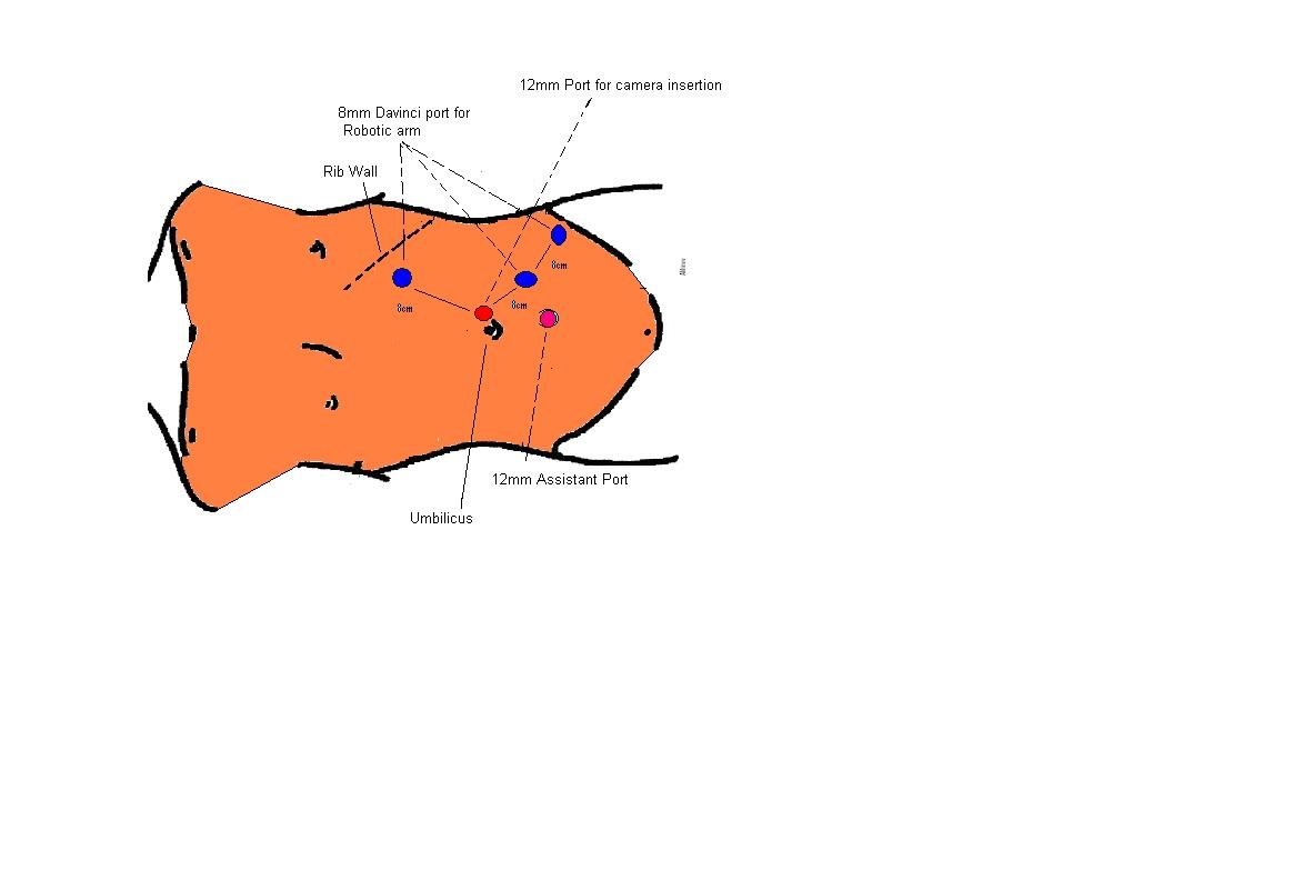
Surgical Instruments
1. Da Vinci Xi Surgical System
2. 0 and 30 deg telescope
3. Meta Clip- P
4. Bipolar graspers
5. Laparoscopic clip appliers
6. Specimen retrieval bag
7. Dextrous Hand Port
8. AirSeal Port
9. Conved Airseal insuffulator
10. Aortic punch
