Laparoscopic APPENDIX
Surgeon(s)/Speaker(s): Dr. Ramesh Makam
Surgical Procedure: Laparoscopic Appendicectomy-Appendix
Location: Vikram Hospital Bangalore
Indexsteps
1. Diagnostic laparoscopy : Diagnostic laparoscopy to exclude any trocar-related injury and un-anticipated pathology.
2. Examination of the caecum / terminal : Examination of the caecum / terminal one meter of small bowel / mesentery / uterus / ovaries and tubes (in females with RIF pain).
3. Freeing up of the omentum : Freeing up of the omentum in cases with acute appendicitis.
4. Identify location : Identify location of the appendix
5. Tackling the Mesoappendix : Appendix is held by its tip with left 5 mm grasper using short bursts of monopolar currents:New:Mesoappendix is dissected free from the organ baring appendix to the base:New:If mesoappendix is very thick or it is inflamed, a bipolar current is advisable (at the base):New:Bipolar current is applied till the boiling stops and the tissue looks white:New: If monopolar current is used, short intermittent bursts given to bare the appendix:New:At the caecal junction, care is taken to avoid thermal injury to the caecum:New:Using 2 atraumatic graspers, caecum is lifted, tinea are traced down to locate the appendix:New:Very often, appendix is visible non adherent, pelvic positioned and pops up on lifting the caecum:New:In case of retrocaecal appendix, caecum is grasped and lateral and inferior peritoneal incisions are made:New:In extensive adhesions, inflamed thick tumour like appendix, harmonic scalpel may be used.
6. Tackling thebase : The base of the appendix once dissected, is ligated with endoloops using 1-0 vicryl, 2 loops on patient side and 1 loop on specimen side.
7. Retrieval of the specimen : Retrieval bag is used to avoid contamination, specimen is retrieved either through the 10mm umbilical incision or extending one 5 mm port site.
8. Re-inspection, drains and closure : After specimen retrieval, base is inspected for bleeding and loop positions are reassured:New:Small bowel walk is done to see for lymph nodes, meckel’s diverticulum (Note the pressure and do not tackle if not pathological):New:Peritoneal cavity cleaned with suction irrigation canula:New:Drains are usually not necessary but in case of contamination or perforated appendix , a drain can be kept:New:All ports are closed, the 10 mm site sheath with port closure needle.
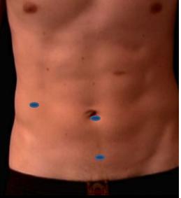
port positions
1. Port Position for Lap Appendicectomy : * Supra-umbilical 10mm trocar (camera port, inserted with closed technique using a Verres needle + sharp trocar or by open method)
* Left iliac fossa 5mm trocar
* Suprapubic 5mm trocar taking care of the bladder
precautionary_measures
1. Retrocaecal appendix : *Caecum is elevated and dissected upwards, even ascending colon needs to be dissected
2. Elongated Retrocaecal appendix : * Dealt in a reverse way, dealing the base first with intracorporeal ligation and later dissecting the mesoappendix
3. Unexposed tip : *Done by tackling the base first and then dissect towards the tip by dividing the mesoappendix close to the appendix
4. Necrotic appendix : Needs careful dissection, if needed intra corporeal suturing done to close caecum
pre_post_measures
1. Preoperative investigations : CBC:new:Chest X-ray,ECG:new:Routine urine
2. Preoperative measures : Fasting for 8 hours before surgery:new: IV to administer fluids, antibiotics and pain medication as prescribed by the surgeon
3. Postoperative measures : Clear liquids the next day after surgery:new:Intravenous antibiotics for 48 hours, converted to oral antibiotics for 5 days:new:Solid food a few hours after that:new:IV removed after patient is on solid food
4. Warning signs in the postoperative period : In case patient develops distension, USG should be performed to detect collection, leak
Surgical Instruments
1. 10mm Trocar
2. Reducer (10mm to 5 mm)
3. 5 mm Trocar
4. Atraumatic grasper forceps
5. 5mm scissor
6. 5mm 0 degree telescope
7. Monopolar/ Bipolar cautery device
8. Harmonic Scalpel
9. 1-0 vicryl
10. Port closure needle
11. Suction Irrigation canula
