BANDED SLEEVE GASTRECTOMY
Surgeon(s)/Speaker(s): Dr. Ramesh Makam
Surgical Procedure: Bariatric-Lap Banded Sleeve
Location: Vikram Hospital Bangalore
Indexsteps
1. Position and Placement : The patient is supine on a dedicated bariatric surgical table with supports for the feet and straps to secure the patient as often the surgeon needs a rather steep head up position to get access to the upper stomach. intermittent pneumatic compression devices are in place along with a patient warming blanket. Two image monitors are placed at the level of the patient's shoulder on either side for uninterrupted viewing by the surgeon and his assistant. Once the anesthetic team have gained good vascular access with the monitoring devices in place the surgical team comes into position, the surgeon often stands on the right side of the patient with the assistant on the left side. The camera person standing the right of the surgeon and the scrub nurse is positioned next to the camera person or next to the assistant surgeon on the left side of the patient. It is always worthwhile to check the various energy settings, insufflator settings, the necessary devices, staplers and cartridges availability before starting the surgery. The surgeon has to have a constant dialogue with the anesthetist, assistant and the scrub nurse especially during positioning and changing the gastric calibration bougies, introduction and with drawl of long bariatric instruments and staplers so that technical errors and collateral damage is minimized.
2. Gastric decompression : A distended stomach (fluid and air) poses technical difficulties in grasping and handling the stomach. Hence good gastric decompression is needed to ensure a easy and safe dissection. The stomach can be decompressed either with an orogastric ryle's tube or by using the side channel of a gastric calibration bougie.
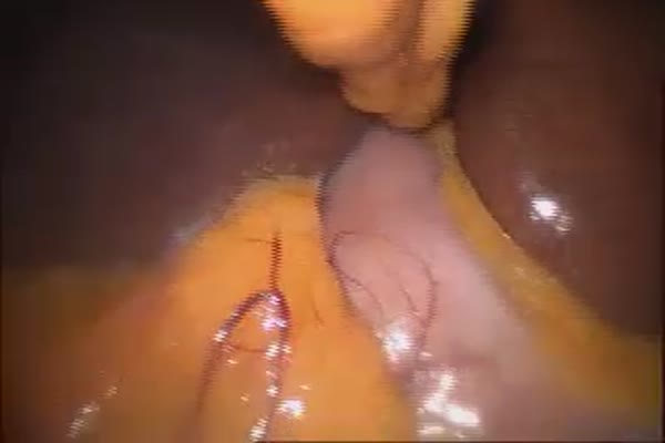
3. Mobilization of greater curvature : The greater omentum is taken down using energy device (harmonic shear, ultrasscion etc) starting at a point about 4cms-6 cms proximal to the pylorus. the dissection is carried out cephalad and the gastric fundus is mobilized in its entirety taking adequate care to safe guard the spleen especially when there are omental and ligamentous adhesions.
4. Creation of the gastric tube : A 32-36 Fr gastric bougie is placed by the anesthetist and is guided by the surgeon along the lesser curve until its down the pylorus. The gastric stapling device (endo GIA) is introduced and the stomach is stapled starting from the point about 4-6 cm from the pylorus. Sequential stapling is done in the cephalad direction until the entire stomach is divided. Care is taken to avoid injury to the pancreas and spleen; staple position and sleeve size is frequently checked to avoid a tight or lax sleeve and to avoid a spiraling staple line due to uneven incorporation of the anterior and posterior gastric walls.
5. Assessing staple line integrity : The bougie is gently withdrawn proximally and air insufflation is done so that the surgeon can check for air leaks by instilling saline over the stapling line. At times a persistent ooze is encountered at specific points along the staple line that needs to be addressed by applying a ligaclip rather than using energy sources as the staples conduct the energy into the deeper tissues. There also exists the practice of placing a layer of surgical wrapping the staple line or re-enforcing the staple line by applying a row of invaginating sutures or by using buttressing materials although this is not standard or mandatory practice.
6. Specimen extraction : The resection stomach is extracted via the umbilical port under vision with the camera being introduced via the 13 mm right sided port. All ports 10 mm and above are closed with vicryl sutures under vision with or without the aid of a port closure device.
Surgical Instruments
1. Harmonic Scalpel
2. Endo Stapler
3. GST Powered Echelon
4. Vessel Sealer